Pancreas
The pancreas is an organ the upper abdomen. Enzymes (chemicals)
made by cells in the pancreas pass into the gut to help digest food. The
hormones insulin and glucagon are also made in the pancreas and help to
regulate the blood sugar level.
What is the pancreas?
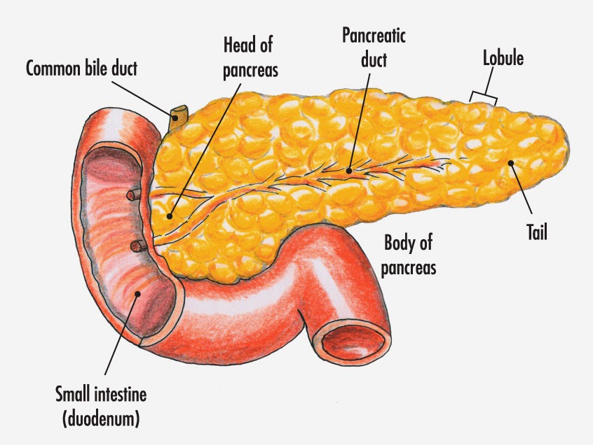
The pancreas is an organ in the
upper abdomen. It is about the size of a hand.
Where is the pancreas found?
The pancreas is in the upper abdomen and lies behind the stomach
and intestines (guts). The pancreas has a connection to the duodenum (the first
part of the gut, which is connected to the stomach) via a duct (tube). This
connecting duct allows the enzymes produced by the pancreas to pass into the
intestines.
What does the pancreas do?
The pancreas has two main functions:
- To make digestive
enzymes which help us to digest food. Enzymes are special chemicals which
help to speed up your body’s processes.
- To make hormones
which regulate our metabolism. Hormones are chemicals which can be
released into the blood-stream. They act as messengers, affecting cells
and tissues in distant parts of your body.
About 90% of the pancreas is dedicated to making digestive
enzymes. Cells called acinar cells within the pancreas produce these enzymes.
The enzymes help to make proteins, fats and carbohydrates smaller. This helps
the intestines to absorb these nutrients. The acinar cells also make a liquid
which creates the right conditions for pancreatic enzymes to work. This is also
known as pancreatic juice. The enzymes made by the pancreas include:
- Pancreatic proteases
(such as trypsin and chymotrypsin) help to digest proteins.
- Pancreatic amylase
which helps to digest carbohydrates (sugars).
- Pancreatic lipase
which helps to digest fat.
Approximately 5% of the pancreas makes hormones which help to
regulate your body’s metabolism. These hormones are made by several different
cells which clump together like little 'islands' (islets) within the pancreas.
The islets are called ‘islets of Langerhans’ and there are about one million
islets dotted about in an adult pancreas. The hormones produced by the cells in
the ‘islets of Langerhans’ within the pancreas include:
- Insulin - which helps
to regulate sugar levels in the blood.
- Glucagon – which
works with insulin to keep blood sugar levels balanced.
- Somatostatin – helps
to control the release of other hormones.
- Gastrin – which aids
digestion in the stomach.
How does the pancreas work?
The
digestive enzymes produced by the pancreas are controlled by the body’s nervous
system and its hormones. When the body senses food in the stomach, electrical
signals are sent via nerves to the pancreas. These signals stimulate the
pancreas to put more enzymes into the pancreatic juice. Acinar cells respond by
increasing the amount of enzymes they produce. The enzymes leave the cells and
pass into tiny ducts (tubes). These ducts join together like branches of a tree
to form the main pancreatic duct. The pancreatic duct drains the enzymes
produced into the duodenum (the part of the gut just after the stomach).
The enzymes are made in an inactive form so that they don’t digest the pancreas itself. Once they enter the intestines the enzymes are activated and can begin breaking food down.
The main hormones released by the pancreas are insulin and glucagon. These hormones help to regulate the amount of sugar found in the blood and the body’s cells. The body’s cells need energy to function. The most readily available form of energy is glucose, a type of sugar. Insulin helps to take glucose from the blood into the cells themselves. This allows the cells to function properly. Glucagon stimulates cells in the liver to release glucose into the blood when levels are low.
The pancreas carefully monitors the level of glucose in the blood. When levels of glucose are high in the blood, cells within the pancreas make insulin. Insulin gets released into the bloodstream where it causes glucose to move into cells. This decreases the amount of glucose in the blood stream, lowering blood sugar levels. Low blood sugar levels stimulate the pancreas to make glucagon. Glucagon works on cells in the liver causing the release of glucose. If sugar levels in the blood rise above normal, the pancreas stops releasing glucagon. Insulin may then be released to balance the system again.
This system helps to keep the level of glucose in your blood at a steady level. When you eat, levels of sugar in your blood rise and insulin helps to bring them down. Between meals, when your sugar levels fall, glucagon helps to keep them up.
Some disorders of the pancreas
Cancer
of the pancreas
There are various types. The common type is called ductal
adenocarcinoma of the pancreas and arises from cells of the pancreatic duct.
This mainly occurs in people over 60. There are some rare types of cancer which
arise from other types of cells within the pancreas. For example, cells in the
pancreas that make insulin or glucagon can become cancerous ('insulinomas' and
'glucagonomas').
Acute
pancreatitis
This is when the pancreas becomes inflamed over a short time -
within a few days or so. It causes abdominal pain. It usually settles in a few
days but sometimes it becomes severe and very serious. The usual causes are
gallstones or drinking a lot of alcohol.
Chronic
pancreatitis
This is when the inflammation in the pancreas is persistent. The
inflammation tends to be less intense than acute pancreatitis but, as it is ongoing,
it can cause scarring and damage. This can cause abdominal pain, poor
digestion, diabetes and other complications. Drinking a lot of alcohol over a
number of years is the common cause.
Type
1 diabetes
With type 1 diabetes the pancreas stops making insulin. It is
treated with insulin injections and a healthy diet.
==--==